Note: movies are in .mov format -- quicktime capability rqd.
VIEW the mitral cell apical tufts.
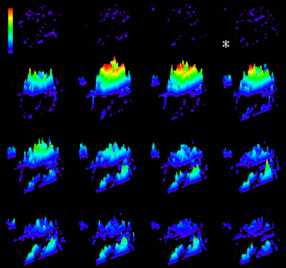
VIEW surface plot movie of the mitral cell tufts.
VIEW surface plot montage of the mitral cell tufts.
Voltage sensitive dye-stained glomeruli
Other images:
Voltage-senstive dye, whole frog brain spontaneous activity
Accessory olfactory bulb terminals
2-Photon Imaging:
eGFP transfected neural crest cells in a chick embryo. 20 hours post transfection, imaged for approx. 1 hour
eGFP transfected neural crest cells in a chick embryo. 20 hours post transfection, imaged for approx. 1 hour
In vivo images of neurons in Line H mice expressing YFP in neurons
In vivo images of olfactory sensory neuron axons and terminals in a mouse expressing tau-eGFP. Mouse courtesy of Jane Roskams, UBC.
Blood vessels in live mouse cortex imaged with fluorescein dextran. In collaboration with Tim Murphy.
See Zhang et al., 2005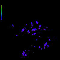